|
PM1 |
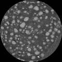
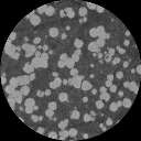
PM2 FOV = 34.2 mm Inter-slice = 74.4 µm |
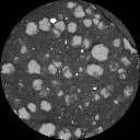
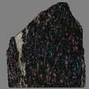
PM4 FOV = 46 mm Inter-slice = 53.4 µm |
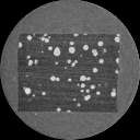
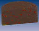
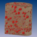
WR1 FOV = 26 mm Inter-slice = 71.9 µm |
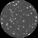
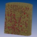
WR3 FOV = 26 mm Inter-slice = 71.9 µm |
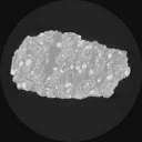
MD FOV = 128 mm Inter-slice = 500 µm |
|
|
Slice Data |
|
 |
 |
 |
 |
 |
|
|
256x256; 3.7 MB 512x512; 11.8 MB |
256x256; 1.9 MB 512x512; 8.0 MB |
256x256; 2.5 MB 512x512; 9.1 MB |
256x256; 2.6 MB 512x512; 9.0 MB |
256x256; 0.7 MB 512x512; 2.2 MB |
|
| 3D Visualization | 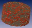 |
 |
 |
 |
 |
 |
| 800x700; 18.1 MB Cylinder 11.3 mm high 22 mm diameter |
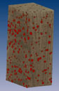
488x740; 17.2 MB Block 1.9 x 2.5 x 4.6 mm |
684x560; 4.6 MB Cylinder 21.4 mm high 46 mm diameter |
600x600; 7.8 MB Cylinder 30.2 mm high 26 mm diameter |
600x600; 7.5 MB Spin: 9.4 MB Cylinder 28.8 mm high 26 mm diameter |
256x256; 2.5 MB 512x512; 7.5 MB Solid ~120 mm high ~120 mm wide |
FOV = CT image field of view; Inter-slice = Distance between CT slice planes.
3D visualizations made using volume rendering, in which each voxel in a data set is assigned a color and an opacity; the latter allows some parts of the specimens to be made transparent, allowing internal features to be viewed.