|
Animation through first CT data set for principal bone-bearing slab.
|
Animation through second CT data set for principal bone-bearing slab |
Animation through CT data set for counter-slab.
|
|
|
|
|
|
|
|
|
SUPPLEMENTAL MATERIAL FOR:
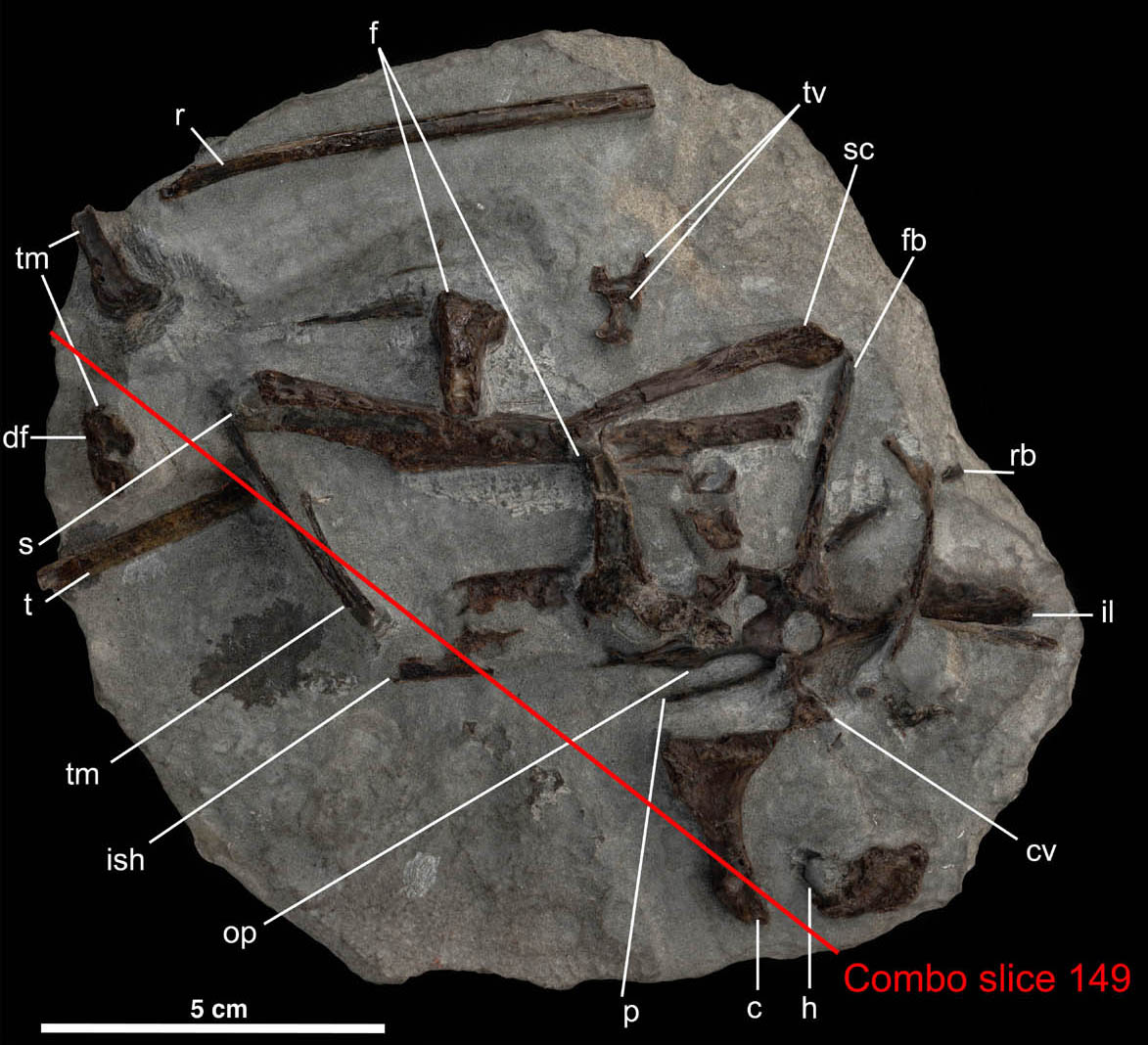
Clarke, J.A., Tambussi, C.P., Noriega, J.I., Erickson, G.M., and Ketcham, R.A. (2005) First definitive fossil evidence for part of the extant avian radiation in the Cretaceous. Nature, 433, 305-308.|
Animation through first CT data set for principal bone-bearing slab.
|
Animation through second CT data set for principal bone-bearing slab |
Animation through CT data set for counter-slab.
|
|
|
|
|
|
|
|
|
CT Methods
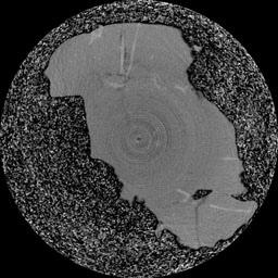
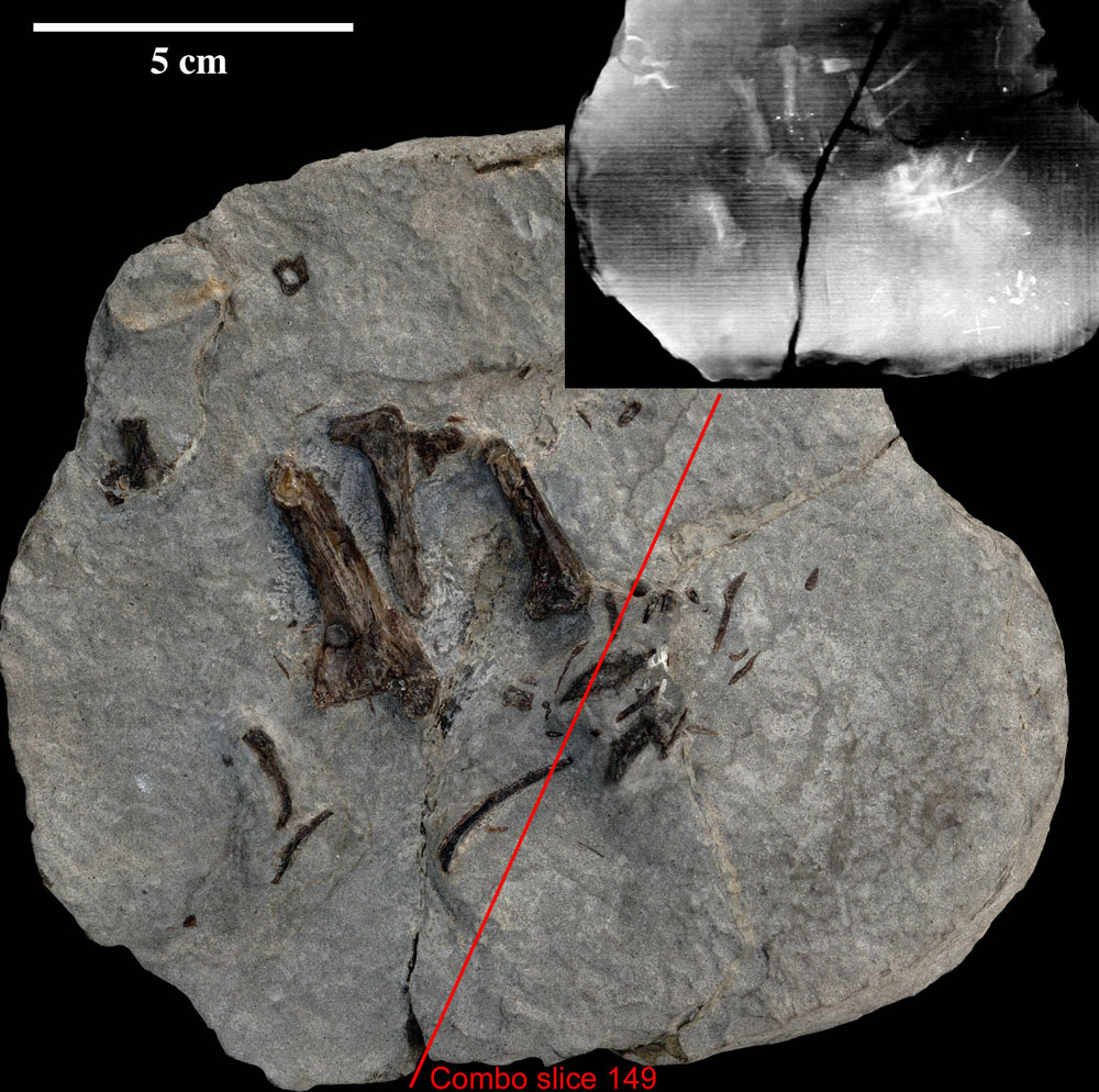
The principal bone-bearing half of the specimen was scanned twice using the high-energy subsystem at the UT CT Facility, using X-ray settings of 420 kV and 4.7 mA. First the specimen was surrounded by garnet sand and scanned horizontally with a slice thickness of 1.0 mm and spacing of 0.5 mm in the principal bone-bearing layer and 1.0 mm below. X-ray settings were 420 kV and 4.7 mA. This protocol provided data relatively free of beam-hardening artifacts but with relatively low resolution. The second scan was done at 0.25 mm slice thickness and spacing with the specimen oriented so that its short axis was parallel to the scan plane. Beam-hardening artifacts, including uneven attenuation values, rings, and streaks, were ameliorated through pre-processing of the raw scan data and post-processing of the reconstructed images. The volume rendering in Fig. 1 was based on this second data set. The counter-slab was also scanned with the second protocol.
Data Processing: Block Realignment
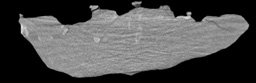
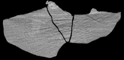
The data sets for each block at 0.25 mm resolution were aligned with each other to reconnect elements that were split along the initial break in the sample. The alignment was done using an algorithm that found the three-dimensional coordinate transform (6-parameter: three translations and three rotations) that maximized the overlap of the margins of the two blocks while not permitting overlap of solid material.
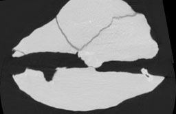
The slice shown below shows the point of contact between the preserved portions of ilium and ishium in both blocks.
 |
 |
 |
|
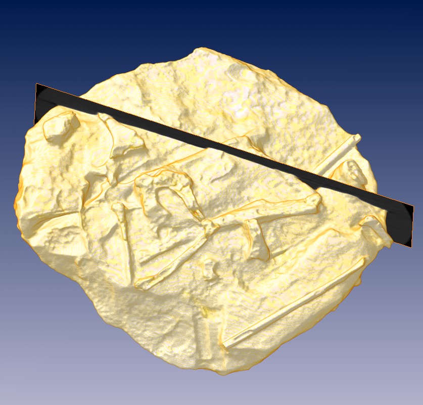
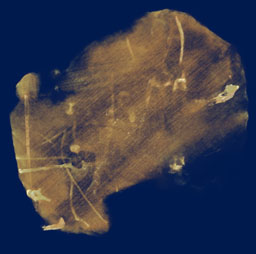
Principal bone-bearing slab, with reference slice from realigned data sets marked.
|
3D reconstruction of principal bone-bearing slab, with reference slice marked.
|
Animations through aligned data
High contrast: 512x332 pixels, 9.2 MB Low contrast: 512x332 pixels; 8.7 MB |
 |
 |
 |
|
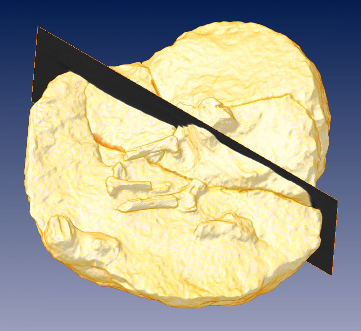
Counter-slab, with reference slice marked.
|
3D reconstruction of counter-slab, with reference slice marked.
|
"Thick-slice" animation through 3D reconstruction of realigned data sets.
1024x864 pixels; 13.9 MB |
![]()